


AI Significantly Improves Pelvic Anatomy Recognition Accuracy, Study Finds
Artificial intelligence can greatly enhance the accuracy of pelvic anatomy recognition, a new study shows, possibly helping surgeons identify critical structures and improve patient safety.
Instead, the clinical language of “image analysis” and “anatomical landmarks” conceals a human story of a deep change in the operating room that reflects the silent but high-stakes anxiety that each and every surgeon carries with them when they operate on the human body.
When a study demonstrates that artificial intelligence can map complex pelvic structures in real time, it is more than just a technical feat. It is a life-saving layer of reassurance for both doctors and patients.
1. Unseen Borders
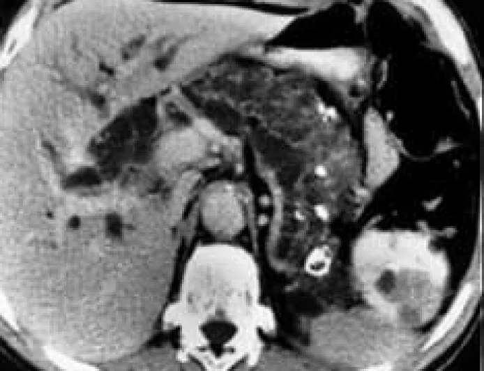
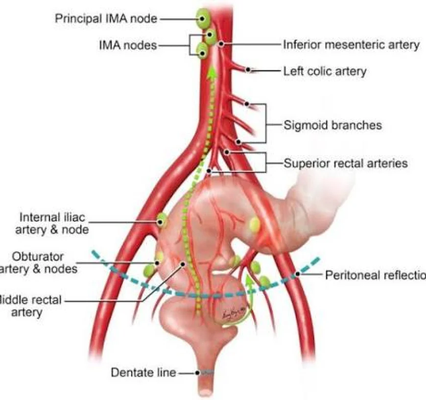
The lower abdomen is a notoriously unforgiving territory for surgeons working on the human pelvis, whether they are clearing out cancer or removing deep lymph nodes. It is a tight, shadowy space, packed close, where vital blood vessels, fragile pathways and fine webs of nerves sit millimetres apart.
The human cost of one misstep here is devastating and could mean permanent nerve damage, severe internal bleeding or loss of organ function. You have to be really focused, because in a long and tiring operation you have to be able to tell the difference between a small, pale nerve and the tissue around it. This important surgical terrain is so challenging that medical literature often describes the ideal borders between tissue layers as the "holy plane", a clear road map that is unbelievably hard to visualise with the naked eye alone.
2. Operating Room Digital GPS
Researchers trained a deep-learning AI model to be an immediate, digital navigator by analysing over 23,000 real-world surgical video frames. The system follows the movement of surgical tools and displays real-time maps directly on the monitor screen during a live procedure.
The clinical advance is in the quality of AI to find important boundaries. It outlines structures like the delicate ureter, the obturator nerve and major iliac arteries with incredible precision, charting the exact safe zones for the surgeon’s scalpel. It doesn’t take over or automate the surgery, but it acts as an unblinking, expert co-pilot that is constantly double-checking the surgical field, catching subtle details that human eyes might miss during a gruelling multi-hour operation.
3. Democratisation of surgical expertise
Perhaps the most profound impact on humans from this technology is that it flattens the learning curve Historically, the art of complex pelvic surgery has required decades of hands-on experience to develop an intuitive, mental library of anatomical variations.
When used in various medical fields, including urology, gynaecology and colorectal surgery, the AI assistance led to an immediate and significant increase in the accuracy of doctors at all career levels. Creating a real-time visual guide for rotating residents and junior fellows makes a stressful educational experience structured and highly predictable. By removing the guesswork from complex anatomy, the technology ensures that a patient’s safety is less dependent on the individual surgeon’s years of tenure and more dependent on a standardised, universally accessible standard of care.
The Symbiotic Future: This is a major paradigm shift in how we think about machine learning in medicine. The technology isn’t trying to replace the irreplaceable touch and judgement of a human specialist but is stepping into a supportive role as a secondary, digital eye that reduces cognitive fatigue and gives doctors the ultimate confidence to operate safely.
What's Your Reaction?
 Like
0
Like
0
 Dislike
0
Dislike
0
 Love
0
Love
0
 Funny
0
Funny
0
 Angry
0
Angry
0
 Sad
0
Sad
0
 Wow
0
Wow
0